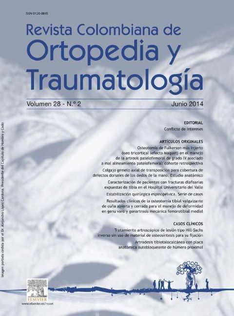
Spinopelvic surgical stabilization: a cases series
DOI:
https://doi.org/10.1016/j.rccot.2015.02.002Keywords:
lumbosacral dislocation fractures, lumbopelvic dissociation, evidence level: IVAbstract
Background: Among the instrumentations that are performed on the spinal column, those of the cranial-cervical and spinopelvic region are the most complex stabilization methods used, given the biomechanical characteristics of these anatomical areas.
Material and methods: A descriptive study of a sequential cases series is presented, which reviews the experience of the patients treated in the period 1992-2012, using spinal column to iliac or sacral (S1 and/or S2) fixations. An evaluation was performed using the demographics, clinical-surgical results according to indications, medical diagnoses, fixation methods used, anatomical extension of the construction in the spinopelvic segment, and complications associated with the technique.
Results: A total of 169 spinopelvic instrumentations were performed on 103 men and 66 women, with a mean age of 37.4 years. The majority of the fixations were performed on the sacrum (153 cases [90.5%]) in patients with spondylitis, lumbar spinal narrowing, and spondylolisthesis. Less frequently were fixations to the iliac (14 cases [8.3%]) in patients with deformities, tumors, trauma, infections, and revision surgery. As complications, in order of frequency, were infections at the surgical site, material failures, or specific complications inherent to the underlying disease.
Discussion: The results were similar to those found in the international literature reviewed as regards surgical indications, instrumentation techniques used, and post-surgical complications. In accordance with the case series analyzed, sacral (S1 and/or S2) fixation is recommended in degenerative disease (spondylolisthesis, spondylolysis, lumbar spinal narrowing), and iliac fixation (conventional Galveston, modular Galveston) in deformities, high energy trauma (lumbopelvic dissociations, L5-S1 inveterate dislocation fractures), and revision degenerative spine surgery (pseudoarthrosis, failure of osteosynthesis material, flat back). In diseases that selectively affect the lumbopelvic area (tumors, infections) sacral or iliac fixation must be chosen depending on the specific anatomical compromise.
Downloads
References
Kasten MD, Rao LA, Priest B. Long-term results of iliac wing fixation below extensive fusions in ambulatory adult patients with spinal disorders. J Spinal Disord Tech. 2010 Oct;23(7):e37-42. https://doi.org/10.1097/BSD.0b013e3181cc8e7f
Matta JE, Ortiz M, Molina MJ, Gamarra RF. Fijación interna de la articulación sacroiliaca inestable. Experiencia Hospital Militar Central, 8 años. Serie de casos. Mejor trabajo de Ingreso, 45. Congreso SCCOT, Cali, agosto 2000.
Bellabarba C, Schildhauer TA, Nork SE, Barei DP, Routt MLC Jr, Chapman JR. Decompression and lumbopelvic fixation for highly displaced sacral fracture-dislocations with neurologic deficits. Spine J. 2004;4:S3-119. https://doi.org/10.1016/j.spinee.2004.05.044
Bellabarba C, Schildhauer TA, Vaccaro AR, Chapman JR. Complications associated with surgical stabilization of highgrade sacral fracture dislocations with spino-pelvic instability. Spine. 2006;15;31:S80-8. https://doi.org/10.1097/01.brs.0000217949.31762.be
Guo-Qing T, Ji-liang H, Bai-Sheng F, et al. Lumbopelvic fixation for multiplanar sacral fractures with spinopelvic instability. Injury. 2012 Aug;43(8):1318-25. https://doi.org/10.1016/j.injury.2012.05.003
Matta JE, Fergusson A, Salamanca JH. Diseño y modificación de técnicas del esqueleto axil. Instrumentación analítica -Investigación Básica Rev Colomb Ortop Traumatol, 9 (1995), pp. 27-35
Matta JE, Rodriguez JM, Ochoa AG, Alvarado GC, Matamoros CH, Rojas VG. Diseño y evaluación clínica de las técnicas de fijación interna modificadas del esqueleto axial instrumentación analítica. Rev Colomb Ortop Traumatol, 9 (1995), pp. 37-48
Matta JE. Introducción al análisis de artículos científicos. Rev Colomb Ortop Traumatol, 10 (1996), pp. 179-182
Van Royen BJ, Van Dijk M, Van Oostveen DPH, et al. The flying buttress construct for posterior spinopelvic fixation: a technical note. Disponible en: www.scoliosisJuornal.com/BiomedCentral.Open
Schildhauer TA, Bellabarba C, Nork SE, Barei DP, Routt ML Jr, Chapman JR. Decompression and lumbopelvic fixation for sacral fracture-dislocations with spino-pelvic dissociation. J Orthop Trauma. 2006 Jul;20(7):447-57. https://doi.org/10.1097/00005131-200608000-00001
Mindea SA, Chinthakunta S, Moldavsky M, Gudipally M, Khalil S. Biomechanical comparison of spinopelvic reconstruction techniques in the setting of total sacrectomy. Spine (Phila Pa 1976). 2012 Dec 15;37(26):E1622-7. https://doi.org/10.1097/BRS.0b013e31827619d3
Gottfried ON, Daubs MD, Patel AA, Dailey AT, Brodke DS. Spinopelvic parameters in postfusion flatback deformity patients. Spine J. 2009 Aug;9(8):639-47. doi: 10.1016/j.spinee.2009.04.008.
Gardocki RJ, Watkins RG, Williams LA. Measurements of lumbopelvic lordosis using the pelvic radius technique as it correlates with sagittal spinal balance and sacral translation. Spine J. 2002 Nov-Dec;2(6):421-9. https://doi.org/10.1016/S1529-9430(02)00426-6
Neubauer P, Skolasky R Jr., Kebaish K. Sacropelvic fixation using the transilial bar technique in adult spinal deformity. Spine. 2008;8:S1-191. https://doi.org/10.1016/j.spinee.2008.06.708
Lehman RA Jr, Kang DG, Bellabarba C. A new classification for complex lumbosacral injuries. Spine J. 2012 Jul;12(7):612-28. https://doi.org/10.1016/j.spinee.2012.01.009
Zahi R, Thévenin-Lemoine C, Rogier A, et al. The "T-construct" for spinopelvic fixation in neuromuscular spinal deformities. Preliminary results of a prospective series of 15 patients. Childs Nerv Syst. 2011;27(11):1931-5. https://doi.org/10.1007/s00381-011-1411-3
Hyun SJ, Rhim SC, Kim YJ, Kim YB. A mid-term follow-up result of spinopelvic fixation using iliac screws for lumbosacral fusion. J Korean Neurosurg Soc. 2010 Oct;48(4):347-53. https://doi.org/10.3340/jkns.2010.48.4.347
Kang DG, Cody JP, Lehman RA Jr. Combat-related lumbopelvic dissociation treated with L4 to ilium posterior fusion. Jr. Spine J. 2012;12:860–861
Ayoub MA. Displaced spinopelvic dissociation with sacral cauda equina syndrome: outcome of surgical decompression with a preliminary management algorithm. Eur Spine J. 2012 Sep;21(9):1815-25. https://doi.org/10.1007/s00586-012-2406-9
Roussouly P, Pinheiro-Franco JL. Biomechanical analysis of the spino-pelvic organization and adaptation in pathology. Eur Spine J. 2011 Sep;20 Suppl 5(Suppl 5):609-18. https://doi.org/10.1007/s00586-011-1928-x
Matta J, Rozo FM, Restrepo FS. Fijación traspedicular y fusion-artrodesis circunferencial para el tratamiento de la espondilolistesis lumbosacra de alto grado. Experiencia multicéntrica Rev Colomb Ortop Traumatol. 2004;18.
Soultanis K, Karaliotas GI, Mastrokalos D, Sakellariou VI, Starantzis KA, Soucacos PN. Lumbopelvic fracture-dislocation combined with unstable pelvic ring injury: one stage stabilisation with spinal instrumentation. Injury 2011;42:1179-1183. https://doi.org/10.1016/j.injury.2010.06.002
Matta I, Diaz S, Gamba CE. Fijación traspedicular para el tratamiento de espondilolistesis, espondilolisis y canal lumbar estrecho de la columna lumbosacra. Experiencia multicéntrica 10 años. Rev Colomb Ortop Traumatol. 2002;16.
Yi C, Hak DJ. Traumatic spinopelvic dissociation or U-shaped sacral fracture: a review of the literature. Injury. 2012 Apr;43(4):402-8. https://doi.org/10.1016/j.injury.2010.12.011
Jones CB, Sietsema DL, Hoffmann MF. Can lumbopelvic fixation salvage unstable complex sacral fractures? Clin Orthop Relat Res. 2012 Aug;470(8):2132-41. https://doi.org/10.1007/s11999-012-2273-z
Downloads
Published
How to Cite
Issue
Section
License
Copyright (c) 2024 Revista Colombiana de ortopedia y traumatología

This work is licensed under a Creative Commons Attribution 3.0 Unported License.


